$180.00
As sonographers, it’s easy to get caught up with patients who require immediate attention for non-ultrasound related issues. Even experienced professionals can overlook the fundamentals of physics. However, when we revisit the basics, we can optimize image quality and enhance our understanding of Doppler principles and hemodynamics.
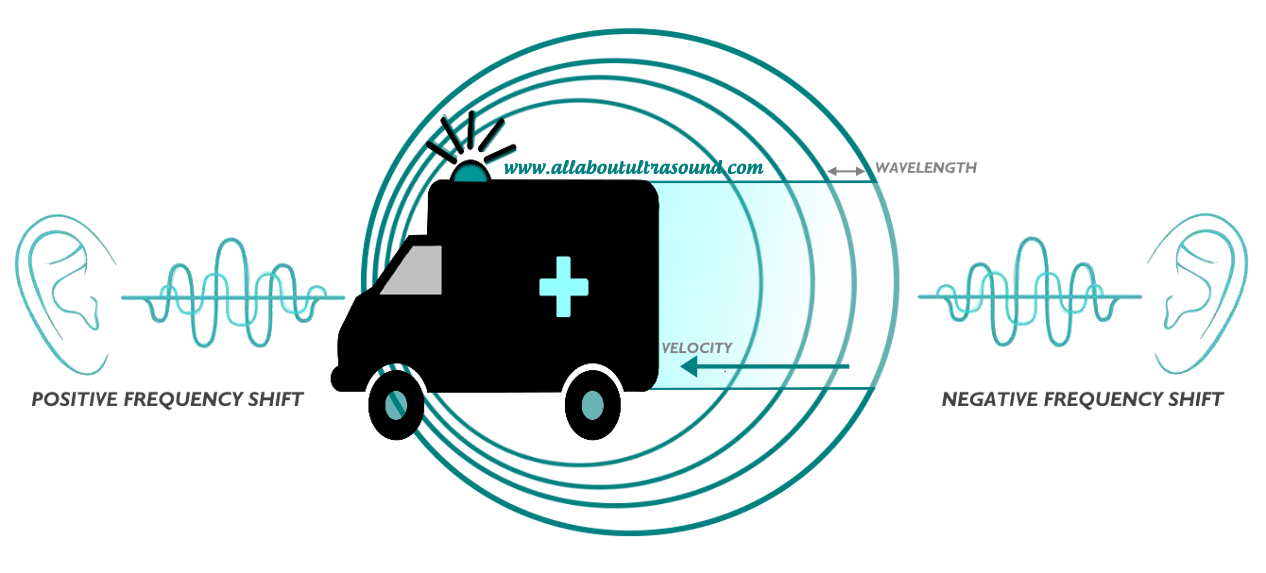
The Doppler Effect is often viewed as a complex concept, but it’s relatively simple when broken down. Essentially, it’s a shift in frequency that occurs when an object moves relative to the observer. This shift can be either positive (compression in wavelength, resulting in a higher frequency) or negative (elongation in wavelength, resulting in a lower frequency).

So, how does the Doppler Effect relate to Doppler ultrasound? The frequency shifts caused by moving red blood cells produce the spectral display we see in Continuous Wave and Pulse Wave Doppler. These velocity measurements can also be displayed in Color Doppler, superimposed on top of the 2D ultrasound image.
In Color Doppler, we see the mean or average velocity, while spectral Doppler display can quantify specific velocities, like peak systolic and end diastolic velocities, at a point in time. This is crucial when placing the Doppler sample and angle of insonation, as the Doppler sample volume is the “observer” and the frequency shift created by the moving red blood cell is what we’re evaluating on the spectral display.
Since the Doppler frequency shift is relative to the position of the observer, it’s essential to consider the angle of insonation. The Society for Vascular Ultrasound recommends maintaining a scanning angle between 45-60 degrees. While we know that angles closer to 0 degrees provide more accurate velocity results due to the math and cosine of the angle, it’s often not attainable in practice, especially during carotid Doppler exams.
Therefore, for reporting and consistency in the lab, it’s best to stay within the range of what can be easily obtained on most exams. By mastering Doppler ultrasound and understanding its fundamental principles, we can improve our image quality and provide more accurate diagnoses.
| Author |
|---|
Fermentum tempor cubilia risus tellus massa dis consectetur dolor.
WhatsApp Chat Oniline